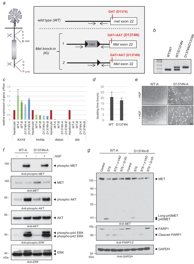
Figure 4. Generation of MET knock-in mice mutated at the C-terminal caspase cleavage site and isolation of hepatic progenitors.
(a) Strategy for generating knock-in mice. The targeted Met allele is depicted. White boxes represent Met exon 22 with, in red, the generated knock-in (KI) mutation GAT >AAT (D1374N). Bold black arrowheads indicate LoxP sites. The positions of the genotyping primers are marked with thin black arrows. (b) To confirm the presence of the KI mutation in Met exon 22, PCR genotyping was performed with primers flanking the loxP sites and amplifying a 476 bp of the wild-type allele (WT) and a 563 bp fragment of the Met KI allele (D1374N). (c) Levels of Krt19, Hnf4a, Aldob, and Alb transcripts were measured by RT-qPCR in Bipotential Mouse Embryonic Liver cell (BMEL) clones derived from WT and MET D1374N mice, and murine hepatocytes as a control. Analyses of two WT BMEL clones (Clones A and B) and two D1374N clones (clones A and B) are shown. The results presented are averages of three independent experiments, with errors bars showing standard deviations. (d) BMEL cells were cultured under routine conditions and were counted after 24, 48, or 72 hr and the doubling times of the WT and MET D1374N BMEL cells were established by averaging the values obtained for the two corresponding clones. (e) Cells were seeded at low confluence. The next day the cells were starved for 30 min in the presence or absence of 10 ng/ml HGF/SF. Representative pictures were taken after 24 hr; scale bar = 100 µm (f) BMEL cells were starved overnight in RPMI-0% FCS and stimulated or not for 10 min with 20 ng/ml HGF/SF. For each condition, the same amount of whole cell lysate was analyzed by western blotting with antibodies against mouse MET, ERK, AKT, and their phosphorylated forms. (g) BMEL cells were treated for 7 hr with 1 µM staurosporine (STS) with or without the pan-caspase inhibitor zVAD-FMK or Q-VD (20 µM). The same amount of protein was resolved by SDS-PAGE and analyzed by immunoblotting with antibodies against the MET kinase domain and cleaved PARP, to assess apoptosis induction, and GAPDH, to assess loading.


