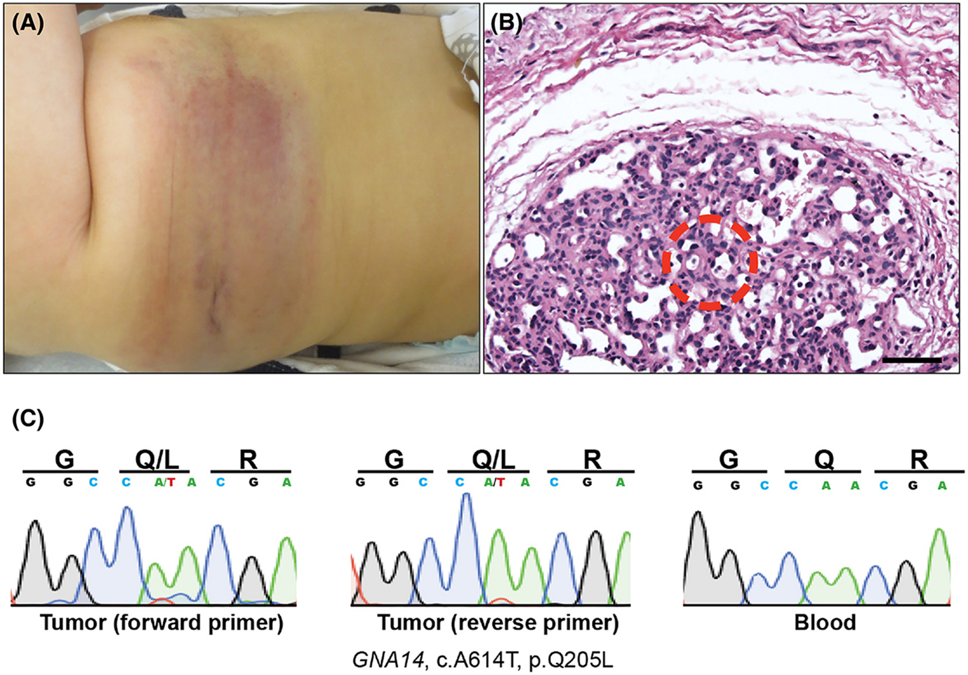
Figure 1. Tufted angioma associated with KMS with an underlying somatic GNA14 mutation.

(A) A violaceous 14×4cm non-blanching patch is observed on the right lateral torso and flank, which was present since birth. (B) Lobular vascular proliferation composed of tightly packed capillaries lined by cytologically-bland endothelial cells (20X magnification, scale bar = 150μm). Area from where LCM was employed to isolated tumor-specific DNA is shown (red circle). (C) Sanger sequencing of tumor (trace data from both primers) and blood control shows GNA14 p.Q205L mutation in tumor only.
