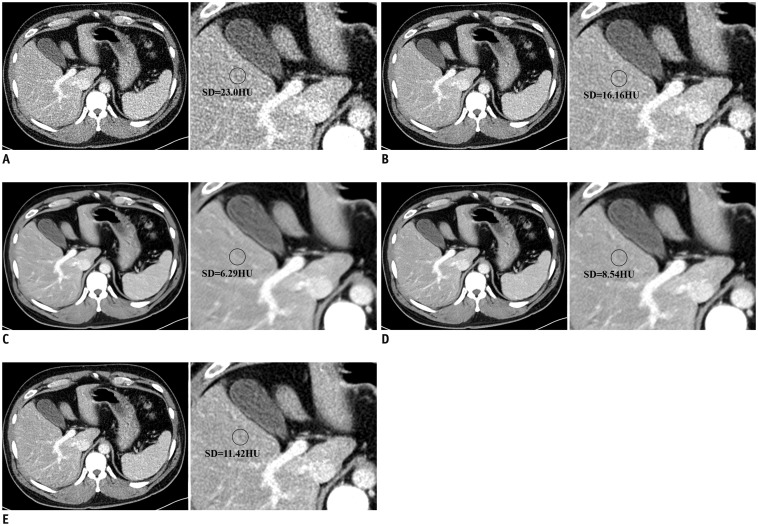
Fig. 4. LD abdominal CT images of test set with conventional reconstruction methods (A, B) and with DLA (C–E).
A. FBP. B. ADMIRE. C. DLA-1. D. DLA-2. E. DLA-3. First column of each image shows LD (25%) abdominal CT using five different methods and enlarged image of first column is in second column. Mean image noise of all LD-DLA images was lower than that of LD-ADMIRE and LD-FBP images, and DLA-1 image showed lowest mean image noise. As training radiation dose of DLA increased, mean image noise of processed CT images increased.

