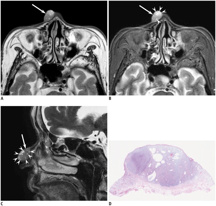
Fig. 1. 87-year-old woman with cutaneous basal cell carcinoma of right nose.
A. Axial T2-weighted image (TR/TE, 3000/90 ms) showing well-demarcated, elliptic, cutaneous lesion (arrow) without superficial ulcer formation and protrusion into subcutaneous tissue. B. Axial fat-suppressed T2-weighted image (TR/TE, 3290/80 ms) showing T2-hyperintense foci (arrowheads) within cutaneous lesion (arrow); peritumoral fat stranding is not observed. C. Sagittal fat-suppressed T2-weighted image (TR/TE, 4350/120 ms) clearly showing T2-hyperintense foci (arrowheads) within cutaneous lesion (arrow). D. Histological specimen (H&E stain, × 2.5) showing well-demarcated mass in dermis with multiple cystic cavities filled with mucinous contents. H&E = hematoxylin and eosin, TE = echo time, TR = repetition time

