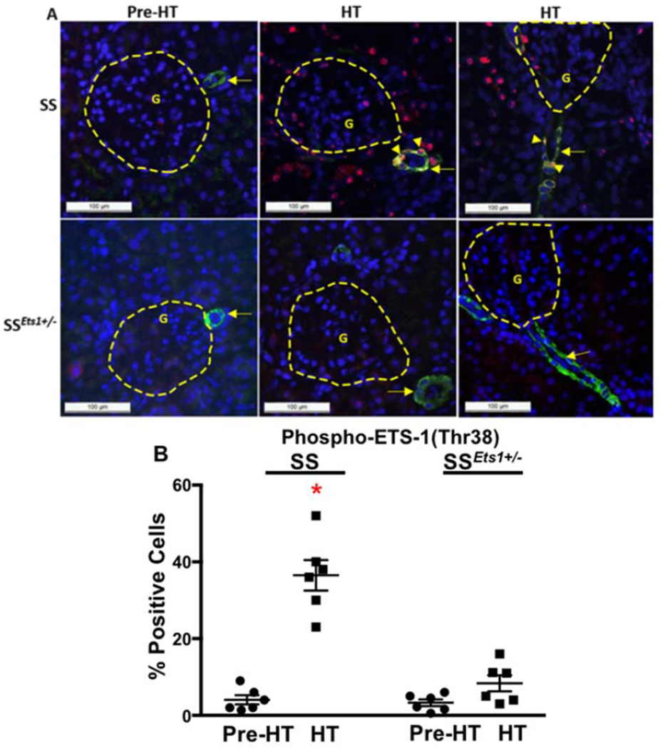
Figure 2. ETS-1 activity increased in kidney microvasculature of HT SS rats, compared with HT SSEts1+/− rats.

A, Immunofluorescence microscopy using alpha-smooth muscle actin (SMA) antibody (Green), which outlined the afferent arteriole, and phospho-ETS-1(T38) antibody (Red), which detected activated ETS-1, was performed in the four groups of rats (Pre-HT SS, HT SS, Pre-HT SSEts1+/−, and HT SSEts1+/−). Basal levels of phospho-ETS-1(T38)-positive cells were low in both Pre-HT SS and SSEts1+/− rats (Upper panel, left). An increase in phospho-ETS-1(T38) staining in the nuclei of smooth muscle cells of the afferent arteriole and microvessels in the kidney of the HT SS rats was observed in both transverse (upper panel, middle) and longitudinal (upper panel, right) views of the afferent arteriole, while the observed numbers of phospho-ETS-1(T38)-positive cells were decreased in the HT SSEts1+/− group. B, Quantification based on determining the ratio of phospho-ETS-1(T38)-positive and -negative cells, which were detected by Alexa Flour 594 staining, in the afferent arteriole showed a dramatic increase in ETS-1 activation in the HT SS group. *p-value < 0.05 versus HT SSEts1+/−, Pre-HT SS and Pre-HT SSEts1+/− groups; n=6 animals in each group. G, glomerulus.
