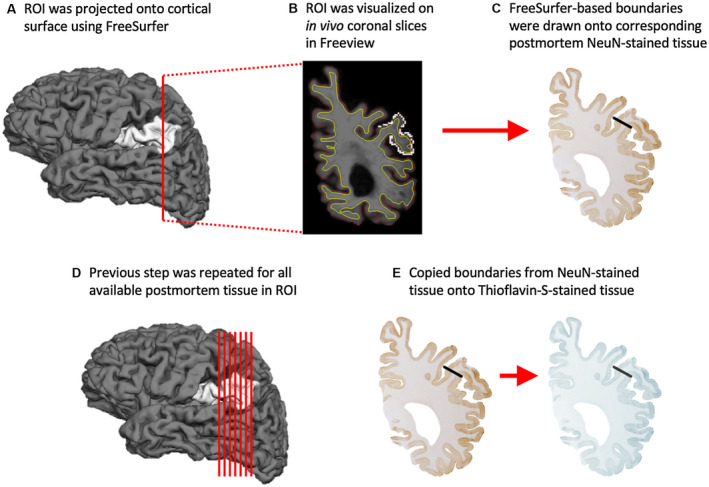
Figure 2.

Anatomic correspondence between in vivo and post‐mortem ROIs. Standardized procedure for matching ROIs used in neuroimaging and post‐mortem analyses. In brief, each FreeSurfer‐generated ROI (A) was initially visualized on high‐resolution MRI scans in the coronal plane (B) before they were matched to every available coronal section of post‐mortem tissue immunohistochemically stained with NeuN (C). The ROI boundaries drawn onto all available post‐mortem NeuN‐positive tissue (D) were then copied onto the parallel series of Thioflavin‐S‐positive tissue (E), restricting stereological quantitation of AD markers to the same anatomical regions where cortical atrophy was measured during life. ROI is the left aIPL.
