Figure 2.
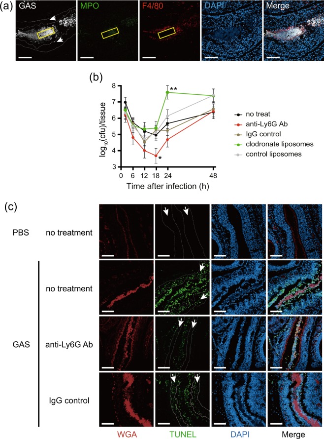
Depletion of neutrophils from mice decreased GAS colonization and damage to the nasal mucosa. (a) Neutrophils accumulated at the local infection site 24 h after nasal infection with the ATCC 11434 strain. Staining with an anti-GAS antibody (white), neutrophil staining with an anti-MPO antibody (green), macrophage staining with an anti-F4/80 antibody (red), and DAPI staining (blue) were performed. The region enclosed by the yellow line is the center of a bacterial aggregate, and the region enclosed by the white dotted line (arrow) represents the epithelial layer. Bars indicate 100 μm. (b) Time-course analysis of the bacterial count in the maxillary sinus after nasal infection with the ATCC 11434 strain. The following 5 groups underwent intranasal infection with 5 × 10⁸ CFU of the ATCC 11434 strain 24 h after the indicated treatments: Intact C57BL/6 mice (no treatment), mice treated with an anti-Ly-6G antibody (anti-Ly6G Ab), mice treated with clodronate liposomes (clodronate liposomes), mice treated with a control IgG antibody (IgG control), and mice treated with control liposomes (control liposomes). The number of live bacteria in the maxillary sinus was counted by seeding specimens on blood agar medium 2, 6, 12, 18, 24, and 48 h after infection. *p < 0.01 (the Student’s t-test, vs. IgG control) **p < 0.01 (the Student’s t-test, vs. control liposomes). (c) Damage to the nasal mucosal epithelium by GAS in mice without neutrophils. Immunostained nasal mucosal epithelial layers 12 h after infection in intact C57BL/6 mice (no treatment), mice without neutrophils using a treatment with an anti-Ly-6G antibody (anti-Ly6G Ab), and mice treated with control IgG (IgG control) infected with 5 × 10⁸ CFU of the ATCC 11434 strain. Samples were stained with WGA (red), TUNEL (green), and DAPI (blue). The region enclosed by the dotted line (arrow) indicates the epithelial layer. Bars indicate 100 μm.

