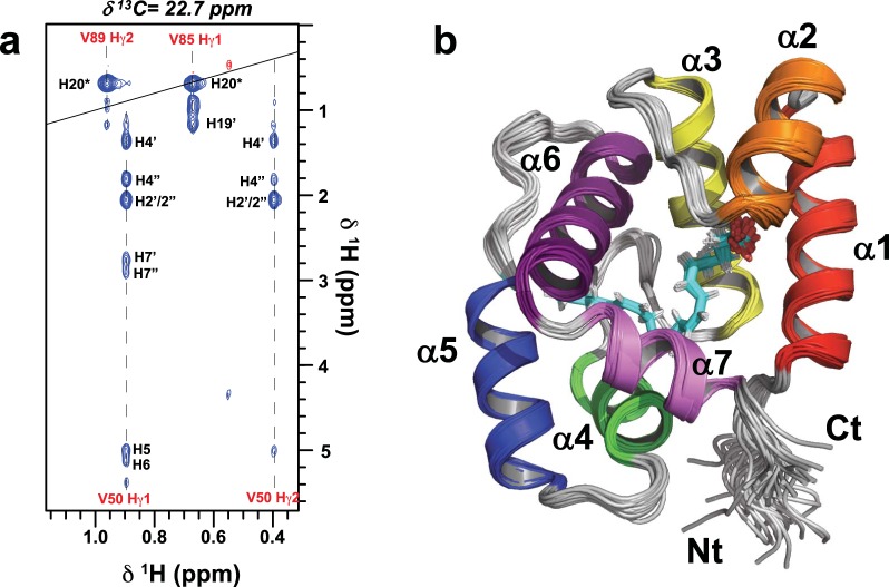
Figure 4.
NMR Structure of the AeOBP22-AA Complex (a) A slice through a 12C-edited/13C-filtered intermolecular NOESY spectrum at δ13C = 22.7 ppm showing NOEs between resonances from the protein (labeled in red) and arachidonic acid (black labels). For the lipid the hydrogens are numbered according to the attached carbon in the alkyl chain. (b) Superposition of the 30 lowest energy structures from the final iteration of NMR calculations (RMSD = 0.32 Å for res 7–121), helices 1–7 are color coded from red-violet. The arachidonic acid is shown in cyan.

