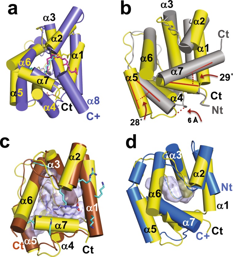
Figure 7.
Comparison of AeOBP22 with known structures. Cylinder representation of AeOBP22 in yellow (N and C-termini labeled in black) compared with (a) N-terminal OBP domain of An. stephensi D7 (blue) bound to Leukotriene C4 magenta (PDB 3NHI)67. For clarity, the C-terminal domain, which continues at the position labeled C+ in blue, is not shown. The linoleic acid bound to AeOBP22 is shown in cyan. (b) LmigOBP1 (grey) (PDB 4PT1)69. The difference in the positions of helices 5 and 7 are shown in red. (c) Phormia regina OBP56a bound to multiple molecules of palmitic acid (blue) (PDB 5DIC) (Ishida et al. not published). The multiple lysines and arginines that surround the pocket (grey) are shown in cyan. (d) An. gambiae OBP22 (bright blue) (PDB 3L4L) (Zhang and Ren, not published). The C-terminus of AgOBP22 is mostly unstructured (extends from position labeled with blue C+).

