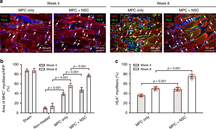
Fig. 6. Myofiber formation and maturation.
a Immunofluorescence for MHC (red)/human leukocyte antigen (HLA, green) of injured TA muscles at 4 and 8 weeks after implantation. The presence of MHC+ HLA+ cells in the implanted region indicates that implanted human MPCs survived and contributed to newly formed muscle fibers (white arrow). b Quantification of the area of MHC+ myofibers per field (%, ×400) (n = 3 per group and time point, four random fields per sample). c Quantification of HLA+ myofibers (%, ×400) (n = 3 per group and time point, three random fields per sample). All data are represented as mean ± SD. The p-values by two-way ANOVA followed by Tukey’s test are indicated.

