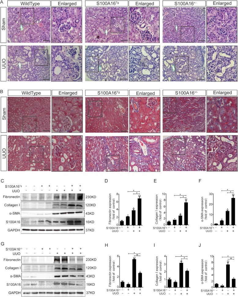
Fig. 2. S100A16 promotes renal interstitial fibrosis in UUO mice.
a, b Representative micrographs of hematoxylin and eosin (HE) and Masson’s trichrome stained kidney tissues demonstrate renal injury in S100A16Tg, S100A16+/−, and wild type mouse kidneys. Scale bar = 50 μm. c–f Representative bands (two cases) of western blots showing the expression of fibronectin, collagen I, and α-SMA in the obstructed kidneys of wild type or S100A16Tg mice. *p < 0.05, **p < 0.01 vs. wild type sham groups; #p < 0.05. g–j Representative bands (two cases) of western blots showing the expression of fibronectin, collagen I, and α-SMA in the obstructed kidneys of wild type or S100A16+/− mice. *p < 0.05, **p < 0.01 vs. wild type sham groups; #p < 0.05, ##p < 0.01.

