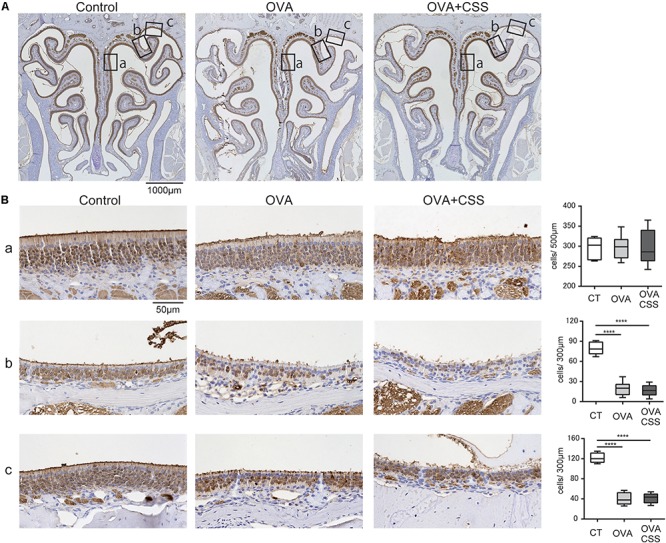
FIGURE 6.

(A,B) Representative immunohistochemical images of olfactory marker protein (OMP)+ mature olfactory receptor neurons (ORNs) in the olfactory epithelium (A) 40× magnification; (B) 400× magnification). Each box (a–c) in (A) indicates the region of the olfactory epithelium shown at a representative higher magnification in (B,a) nasal septum; (b) upper-lateral area; (c) uppermost-lateral area. Comparative charts of cell counts of OMP+ ORNs in each group are shown. ****P < 0.0001 (n = 6, one-way analysis of variance). OVA, ovalbumin; CSS, cigarette smoke solution.
