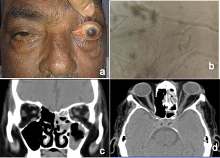
Fig. 1.
a Photograph of patient with left eye proptosis and ophthalmoplegia. b KOH mount showing broad aseptate fungal hyphae with right angled branching. c CT Scan coronal image showing opacification of ethmoid and maxillary sinus on left side. d CT axial image showing proptosis left eye with orbital fat stranding and opacification of ethmoid sinus. There is evidence of ill defined soft tissue density at the orbital apex with bulky left cavernous sinus

