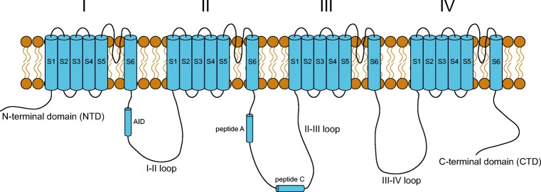
Fig. 2.
Topology of the membrane-embedded pore-forming α1s-subunit of the skeletal muscle dihydropyridine receptor (CaV1.1). Each of the membrane-spanning motifs I–IV is composed of six transmembrane helices S1–S6. The five cytosolic regions of the protein include the N-terminal domain (NTD), I–II loop, II–III loop, III–IV loop and the C-terminal domain (CTD)

