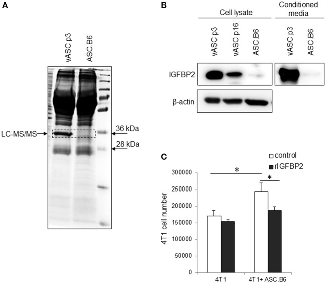
Figure 2.
IGFBP2 expression of vASCs. (A) SDS-PAGE analysis is shown of the proteins precipitated from conditioned media of vASCs or ASC.B6 cell culture, stained with Coomassie Brilliant Blue R-250. (B) IGFBP2 protein was detected by Western blotting experiment from the cell lysate of vASCs at passage number p3 and p16, and ASC.B6 cell cultures or conditioned media of vASCs p3 and ASC.B6 cell cultures. β-actin was used as loading control. (C) Recombinant IGFBP2 (1 μg/ml) inhibited the proliferation of 4T1 cells in the presence of ASC.B6 at a ratio of 2.5:1 in Transwell inserts. The bars show the mean ± SD from three independent experiments, the statistical analysis was t-test with P-values set at: *P < 0.05.

