
Figure 1.
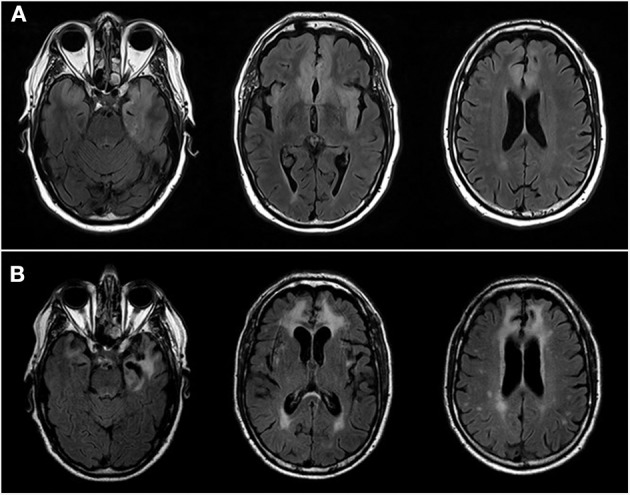
3T Brain MRIs. (A) Patient N°1 MRI at the acute phase of the herpetic meningoencephalitis. Axial images showing hypersignal of the anterior, intern, left, temporal lobe including uncus and hippocampus, left insula and bilateral, ventromedial, orbito-frontal and cingulate anterior lobes. (B) Patient N°1 MRI at the time of AIE. Axial FLAIR images showing sequels of herpetic meningoencephalitis, including extension of previously describe lesions and necrosis of the medial, anterior, left, temporal lobe. Of note, increase of vasculoderenerative subcortical, bilateral lesions.