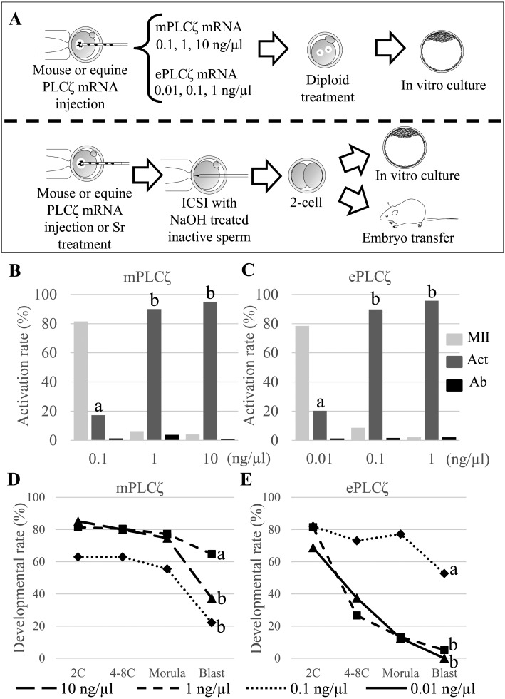
Fig. 1.
Activation and in vitro development of mouse oocytes injected with mouse PLCζ (mPLCζ) or equine PLCζ (ePLCζ) mRNA. (A) Upper; Schematic representation of injection of different concentrations of mPLCζ or ePLCζ mRNA into mouse oocytes. The oocytes were treated with cytochalasin B for 6 h to induce diploid parthenogenesis. Lower: NaOH-treated inactive spermatozoa were injected into oocytes activated by mPLCζ or ePLCζ mRNA injection before culturing in vitro for four days or transferring into a recipient female at the 2-cell stage. (B, C) Rates of activated oocytes after injection of different concentration of mPLCζ or ePLCζ mRNA. The oocytes were injected with 0.1, 1, and 10 ng/µl of mPLCζ mRNA (B), or 0.01, 0.1, and 1 ng/µl of ePLCζ mRNA (C). Experiments were repeated more than six times for each concentration. (D, E) Rates of parthenogenetic embryo development in vitro at different concentrations of mPLCζ or ePLCζ mRNA. Embryos were observed at 24 h (2-cell stage), 48 h (4–8-cell stage), 72 h (morula stage), and 96 h (blastocyst stage). The “a” vs. “b” denotes significant differences between experiments (P < 0.01).

