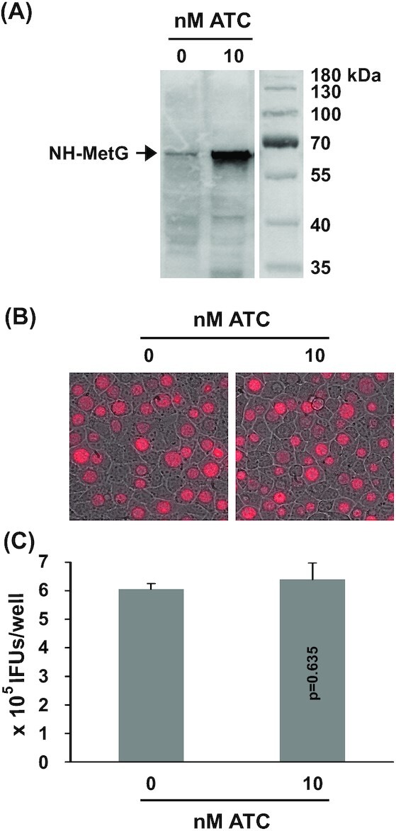
Figure 6.

CtL2 growth unaffected by ATC-induced NH-MetG expression. (A) Western blot detecting the 65 kDa NH-MetG in pTRL2-NH-metG transformants of CtL2 cultured in the presence of 0 or 10 nM ATC. (B) mKate-expressing inclusions formed in the presence of 0 or 10 nM ATC. (C) Recoverable IFUs formed in the presence of 0 or 10 nM ATC. Data are averages ± standard deviations of triplicate experiments. P values are of two-tailed t tests. (A-C) ATC was added at the time of inoculation. Microscopy and determination of recoverable inclusion-forming units were performed at 30 hours post-inoculation.
