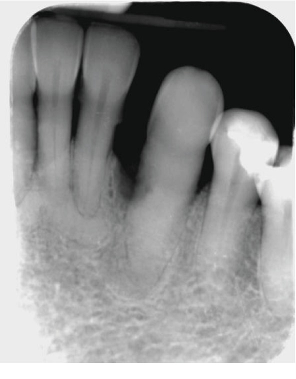
Figure 2.

Long-cone periapical radiograph of the LL3 showing loss of about 50% bony support, obliteration of the root canal, radiolucency on the mesial surface of the root, and sclerosing osteitis periapically. No signs of caries intraorally. The LL2, LL1, and LR1 show 50-70% bone loss and bony sclerosis apically which could indicate late-stage periapical cemento-osseous dysplasia.
