Figure 1.
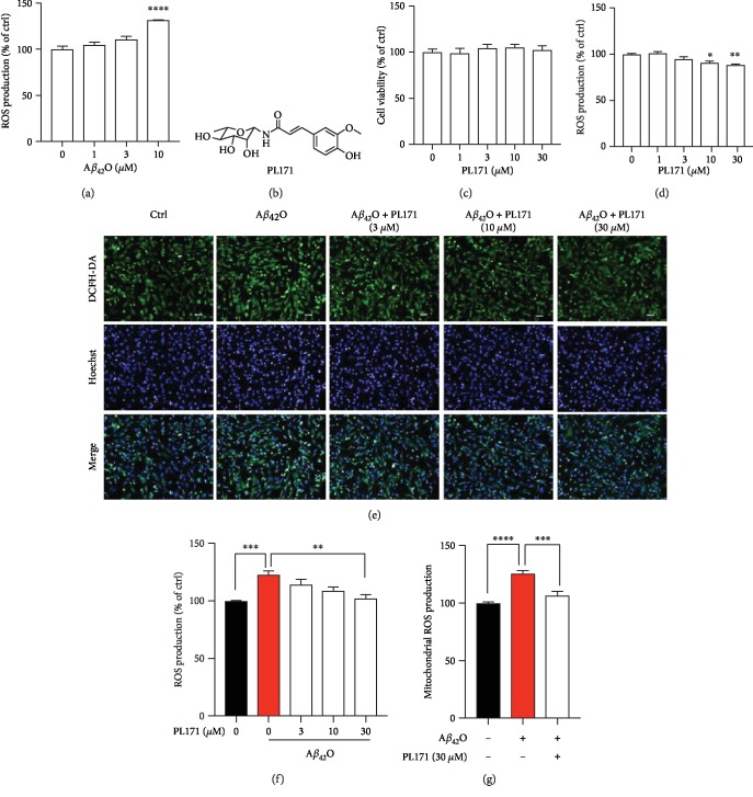
PL171 dose dependently inhibited Aβ42O-induced ROS production in SK-N-SH cells. (a) ROS generation in SK-N-SH cells incubated with Aβ42O (1-10 μM) for 24 h and then stained with DCFH-DA. The data were normalized to the control. (b) The chemical structure of PL171. (c) Cells were treated with PL171 (1-30 μM) for 24 h and cell viability was measured by CellTiter-Glo Assay. (d) The ROS generation of SK-N-SH cells treated with PL171 (1-30 μM) for 24 h followed by staining with DCFH-DA dye. (e) The representative image of SK-N-SH cells preincubated with PL171 (3-30 μM) for 4 h, treated with Aβ42O (10 μM) for another 24 h, and then costained with DCFH-DA and Hoechst. The pictures were obtained by Operetta. Scale bars, 50 μm. (f) The quantification of (e), showing relative ROS generation of cells pretreated with PL171 for 4 h before Aβ42O (10 μM) stimulation for 24 h. (g) Mitochondrial ROS production in the cells challenged of Aβ42O (10 μM, 24 h) with or without PL171 (30 μM, 4 h) preincubation. The signal of MitoSOX was normalized to Hoechst. The data are presented as mean ± SEM, n ≥ 3 independent experiments, ∗p < 0.05, ∗∗p < 0.01, ∗∗∗p < 0.001, and ∗∗∗∗p < 0.0001, analyzed by one-way ANOVA followed by Bonferroni's test.

