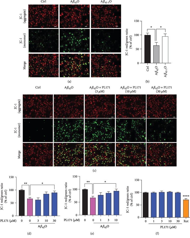
Figure 2.
PL171 prevented Aβ42O-induced MMP reduction in SK-N-SH cells. (a) The representative MMP images of SK-N-SH cells incubated with Aβ42O or Aβ42-1O (10 μM) for 24 h and then stained with JC-1 dye. Green (excitation: 490; emission: 530); red (excitation: 525; emission: 590). Scale bars, 50 μm. (b) The ratio of red/green fluorescence from (a). (c) The representative images of SK-N-SH cells preincubated with PL171 for 4 h followed by treatment with Aβ42O (10 μM) for 24 h. Cells were then stained with JC-1 dye and imaged by Zeiss Observer Z1 microscope. (d) The fluorescence intensity in (c) was quantified using BioTek SynergyNEO. Scale bars, 50 μm. (e) Cells were treated as (d) but with PL171 preincubation for 24 h. (f) Cells were treated with PL171 (1-30 μM) for 24 h, stained with JC-1 dye, and detected by BioTek SynergyNEO. Rotenone (Rot) was the positive control. The data are presented as mean ± SEM, n ≥ 3 independent experiments, ∗p < 0.05, ∗∗p < 0.01, and ∗∗∗∗p < 0.0001, analyzed by one-way ANOVA followed by Bonferroni's test.

