Figure 4.

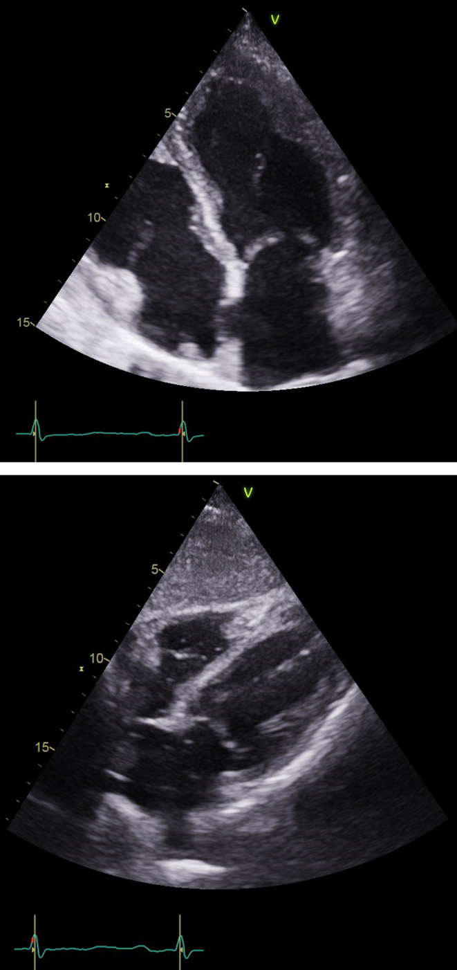
(A) Echo: Subcostal view at the most recent follow-up showing no further evidence of pericardial effusion. (B) Apical four-chamber view showing LV thrombus no longer present.

(A) Echo: Subcostal view at the most recent follow-up showing no further evidence of pericardial effusion. (B) Apical four-chamber view showing LV thrombus no longer present.