
. 2020 Jan 28;7(1):G1–G18. doi: 10.1530/ERP-19-0050
 This work is licensed under a
 Figure 4.
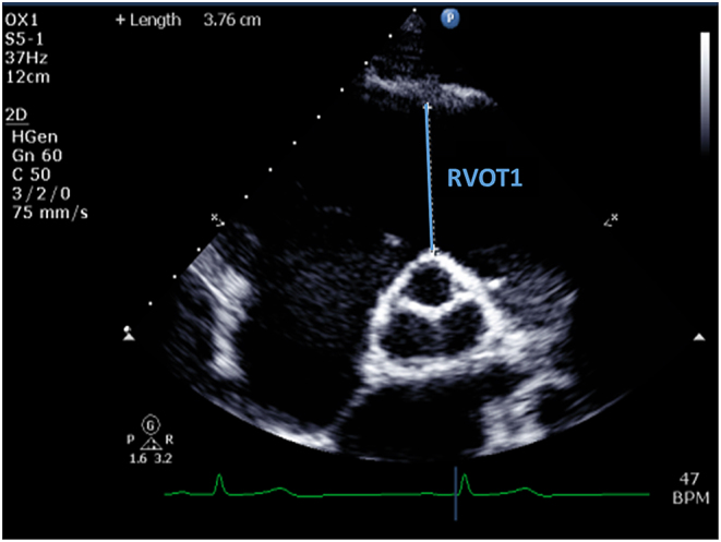
RVOT assessment (RVOT1). From the PSAX window, and in end-diastole, measurement should be made from the anterior aortic wall directly up to the RVOT free wall (at the level of the aortic valve). The PSAX view is more reproducible than RVOT PLAX.