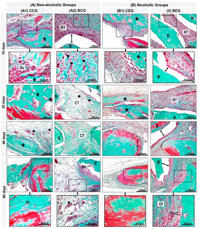
Figure 4.
Details of evolution of bone healing of cranial defects created in the Non-Alcoholic (A) and Alcoholic (B) animals treated with blood clot or biomaterial. (A1) CCG (Non-alcoholic Group; defects filled with blood clot): at 10 days, defects showed trabecular bone formation (asterisk), with the presence of osteoclastic cells (Oc) on the edge of the remaining bone tissue. 20–40 days, the new bone formed showed bone maturation phase surrounded by blood capillaries (black arrow). At 60 days, collagen fibers were arranged in a more regular manner (Cf). (A2) BCG (Non-alcoholic Group: defects filled with biomaterial): at 10 days, defects showed tissue reaction (RT) surrounding the particles of the biomaterial (B); artifacts in histologic sections (double arrow–gap between biomaterial and tissue). Between 20–60 days, connective tissue (CT) presented scarce inflammatory cells with thin and thick collagen fibers, which were parallelly arranged at the end of the experimental period. (B1) CEG (Alcoholic Group; defects filled with blood clot): in the early periods, the defects presented inflammatory cells, decreasing at 40 days. The bone tissue formed at 60 days was predominantly compact and mature. (B2) BEG (Alcoholic Group; defects filled with biomaterial): 10–20 days, sections showed discrete bone formation, and biomaterial particles permeated by reaction tissue. In the later periods, collagen fibers were organized in parallel, and osteoblastic cells (Ob) forming a single cell line adjacent to the matrix. Masson’s Trichrome; original magnification ×40; bar = 100 μm; and Insets, magnified images ×100; bar = 50 μm.

