Figure 4.
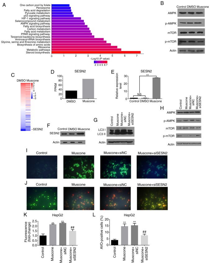
Muscone induces HepG2 cell autophagy through the SESN2/AMPK/mTOR1 signaling pathway. (A) Top Kyoto Encyclopedia of Genes and Genomes (KEGG) pathways enriched in HepG2 cells following treatment with DMSO vs. muscone. (B) Expression levels of AMPK/mTOR signaling pathway proteins in HepG2 cells treated with DMSO or muscone for 24 h were assessed using western blotting. (C) SESN2 gene expression is increased by muscone treatment. (D) FPKM of SESN2 expression identified by RNA-Seq. (E) Relative expression levels of SESN2 in HepG2 cells was assessed using reverse transcription-quantitative PCR following treatment with DMSO or muscone. (F) Expression levels of SESN2 in HepG2 cells treated with DMSO or muscone were assessed using western blotting. (G) LC3 protein expression levels in siSESN2-transfected HepG2 cells treated with muscone for 24 h were assessed using western blotting. (H) Phosphorylation of mTOR and AMPK in HepG2 cells was detected using western blotting in siSESN2-transfected HepG2 cells treated with muscone for 24 h. (I and J) Representative micrographs of (I) MDC staining and (J) AO staining of HepG2 cells at an ×100 magnification. (K) MDC fluorescence intensity fold-change was measured using a fluorescence microplate reader. (L) Positive AO staining cell counting. Densitometric analysis was performed using ImageJ software. **P<0.01 vs. control group; ##P<0.01 vs. muscone group; n=3; NS, not significant; MDC, monodansylcadaverine; AO, acridine orange; LC3, light chain 3; SESN2, anti-sestrin 2; AMPK, AMP-activated protein kinase; mTOR1, mechanistic target of rapamycin kinase 1.

