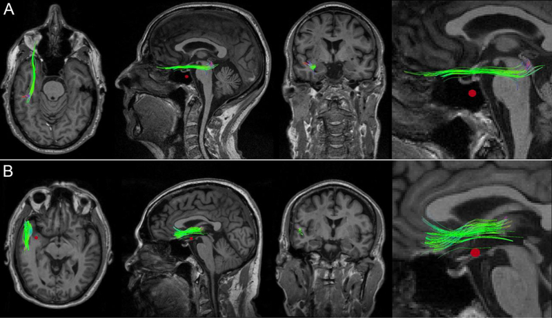
Fig. 3.
WM streamline clusters in two mTBI victims who exhibit a CMB in the WM of the left anterior temporal lobe. In both (A) and (B), FA changes at vertices along the fasciculi were significantly correlated with the distances from these points to the nearest CMB (red). Whereas only some of the WM streamlines in each cluster pass through the CMB penumbra, the observed FA changes were found to extend beyond the immediate spatial neighborhood of the vascular lesion. From left to right: axial view, sagittal view, coronal view, and inset.

