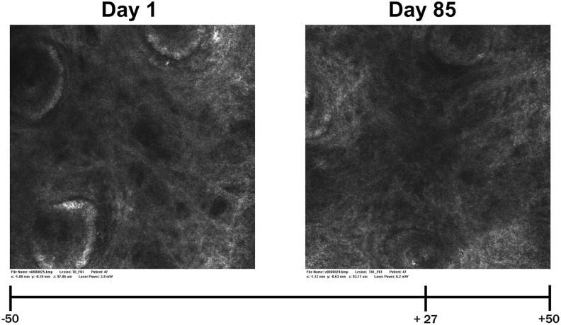
FIG. 3.
VIVASCOPE images (subject RD47) showing the collagen structure of the subcutis, taken before (left) and after (right) 85 days of intake of the test product; VAS at the bottom. The blinded expert grader assessed the right image clearly better than the left image with a score of +27 (0 = no difference). VAS, visual analog scale.

