Figure 5.

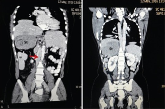
Coronal computed tomography image reveals inferior vena cava (red arrow) of the right lower abdomen continues above with prominent azygos vein (white arrow), right-sided stomach and multiple spleens

Coronal computed tomography image reveals inferior vena cava (red arrow) of the right lower abdomen continues above with prominent azygos vein (white arrow), right-sided stomach and multiple spleens