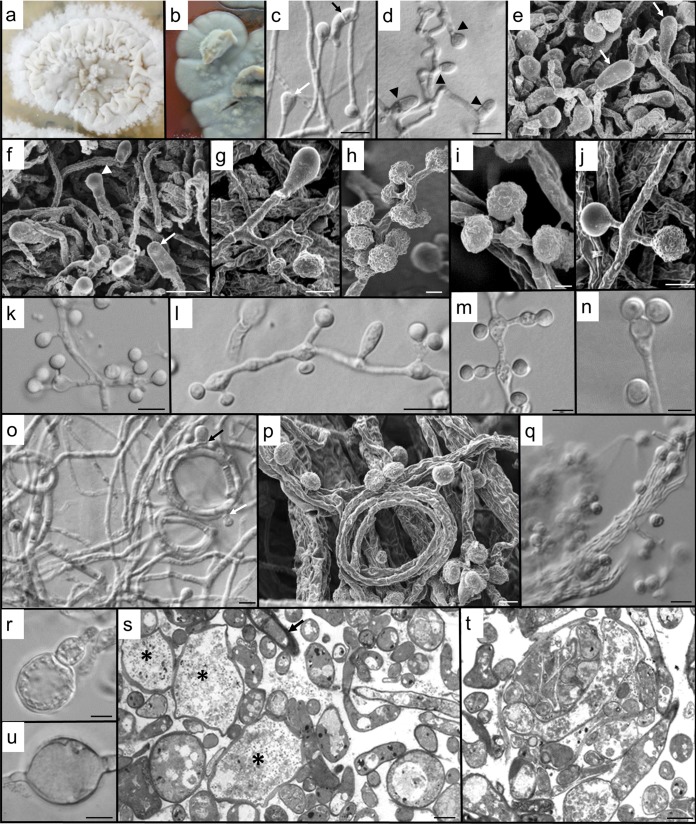
FIG 2.
Morphology of B. emzantsi. Shown are the saprophytic phase (25°C) on Sabouraud agar after 2 weeks (a, c to k, and o to q) and the intermediate phase (30°C) after 3 weeks on BHI agar with 5% horse blood (b) or Sabouraud dextrose agar (l to n and r to u). (a) Floccose, white colony, with a narrow margin peripheral to the crumpled central areas. (b) At 30°C on BHI agar with 5% horse blood, colonies were gray (darker gray on the reverse), with numerous aerial hyphal tufts and a flattened margin. (c) Two terminal conidia, a developing clavate cell (white arrow), and a clavate cell with secondary septation (black arrow). (d) Hyphal septation (arrowheads) in conidiogenesis and clavate cell formation. (e) Clavate, complanate cells (arrows) clearly distinguishable from developing conidia. (f) Complanate terminal cells (arrows) and a developing terminal conidium (arrowhead). (g) Hyphal cell subtending single, lateral conidia and a terminal, basally septate, clavate cell. (h) The apparent clusters of conidia are usually due to the short pedicels of many of the laterally positioned conidia. (i) Multiple conidia on short secondary conidiophores can be subtended from a single basal cell. (j) Conidial ornamentation is variable, from glabrous to papillate. (k) The single conidium may be lateral or clustered on vesiculate primary conidiophores. (l) Clavate cell between conidiophores, with variously arranged conidia. Some inflation of subtending cells is apparent at an intermediate temperature. (m) Increase in temperature results in conidiophores becoming increasingly ampulliform and vesiculate, with distended hyphal cells. Light microscopy creates the illusion that one of each of the laterally paired conidia is sessile. (n) Expanded primary conidiophore with two conidia on extremely short secondary conidiophores. (o) Helical hyphae, one with both developing clavate cells (black arrow) and conidia (white arrow). (p) Numerous conidia developing on hyphal gyres, with bundles of older hyphae behind them. (q) Abundant verrucose conidia and hyphae aggregated into rope-like structures. (r) Development of an adiaspore-like cell (inflated terminal conidia). (s) Sectioned material providing accurate imaging of the diversity of cell forms, including the development of giant cells (*) from enlarged, fragmented hyphal cells; increased vacuolation of enlarging cells; numerous thin hyphal profiles; and some thick-walled cells (arrow) (tangentially sectioned). (t) Section through the hyphal bundles seen by light and scanning microscopy revealing numerous endo/intrahyphal profiles. (u) An inflated intercalary cell suggestive of a chlamydospore. Bars, 5 μm (c, d, k to o, q, s, and u) and 2 μm (e to j, p, r, and t).

