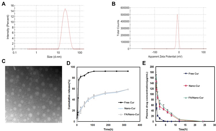
Figure 2.
Characterization of FA/Nano-Cur micelles and drug release study. The particle size and the zeta-potential of FA/Nano-Cur micelles measured by the dynamic light scattering (DLS) method (A and B). Morphological identification of FA/Nano-Cur micelles under transmission electron microscopy (TEM) (C). Cur release from Free Cur, Nano-Cur micelles and FA/Nano-Cur micelles (D). Plasma drug concentration of Cur in Free Cur group, Nano-Cur group and FA/Nano-Cur group at different time point (E).

