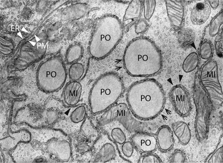
Figure 1.

Electron micrograph of organelle contact sites in the rat liver. The center of the image shows five peroxisomes (PO), which are surrounded by a reticular network of smooth ER tubules (arrows). Furthermore, several mitochondria (MI) with ER MAMs can be found (black arrowheads). Note an elongated mitochondrion (center) in direct apposition to the PO‐ER contacts suggesting the existence of functionally relevant organelle triple contacts. ER—plasma membrane (PM) contacts are also observed (upper left corner; white arrowheads). Magnification: ×25 000 (kindly provided by W. Kriz, University of Heidelberg, GER)
