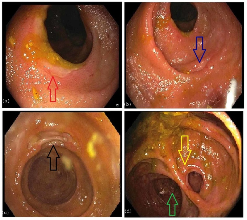
Figure 2. Colonoscopy.
a) depicts ulceration (red arrow) in the ascending colon. b,c) depict ulceration in the distal 15 cm of the neo-terminal ileum (blue and black arrows) with normal-appearing intervening mucosa. d) visualizes anastomosis of the distal ileum (yellow arrow) to the transverse colon (green arrow).

