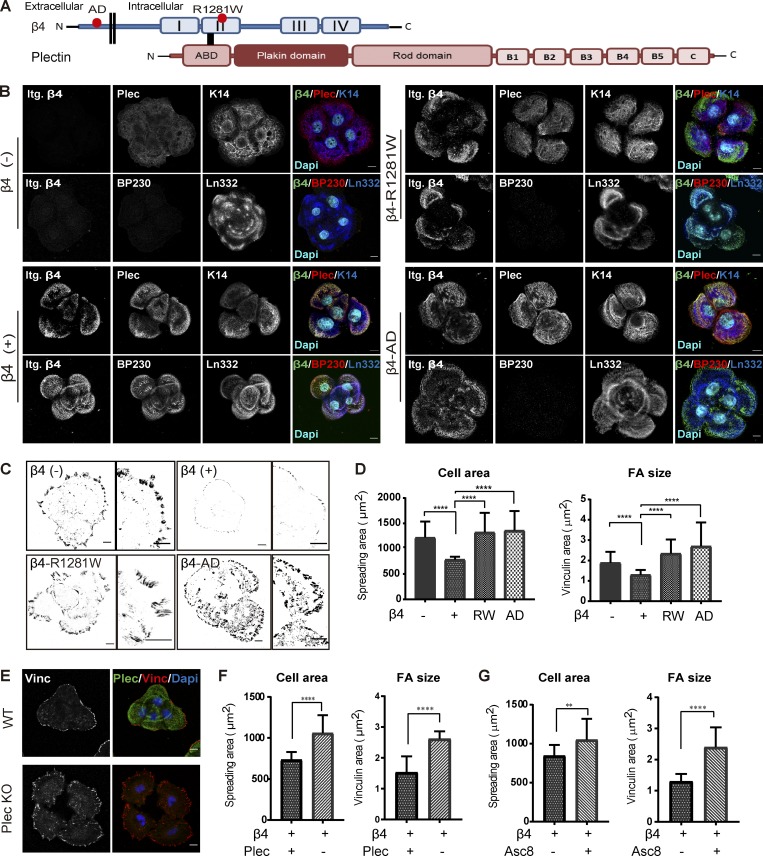
Figure 3.
Intact laminin-integrin β4-plectin linkage reduces FA size and cell spreading. (A) Domain structure of integrin β4 and plectin. Dots indicate the relative locations of the R1281W mutation in β4-R1281W and of the D230A, P232A and E233A mutations in β4-AD. ABD, actin-binding domain. (B) Representative confocal fluorescence microscopy images of β4 (−), β4 (+), β4-R1281W, and β4-AD PA-JEB keratinocytes. Cells were cultured for 24 h in DMEM (10% FCS) and then fixed and stained for β4 (green), plectin (Plec; red) or BP230 (red), and keratin-14 (K14; blue) or laminin-332 (Ln322; blue). Nuclei were counterstained with DAPI (cyan). Scale bars: 10 µm. (C) Inverse black-and-white images of confocal micrographs of β4 (−), β4 (+), β4-R1281W, and β4-AD PA-JEB keratinocytes showing cell morphology and vinculin-stained FAs (black). Scale bars: 10 µm. (D) Quantification of cell area and FA size with ImageJ. The data are presented as the mean (± SD) from three independent experiments, with ∼20 images per experiment. ****, P < 0.0001. (E) Confocal microscopy images of vinculin-stained FAs (red) and plectin-stained HDs (green) in β4 (+) and plectin-deficient β4 (+) keratinocytes (Plec KO). Nuclei (blue) were visualized with DAPI staining. Scale bars: 10 µm. (F) Quantification of cell area and FA size from three independent experiments, with ∼20 images per experiment. ****, P < 0.0001. (G) PA-JEB/β4 keratinocytes were cultured for 24 h in DMEM (10% FCS) with or without the β4 blocking antibody ASC-8 (supernatant diluted 1:5). Shown are quantification of cell area and FA size from three independent experiments, with ∼20 images per experiment. **, P < 0.01; ****, P < 0.0001.

