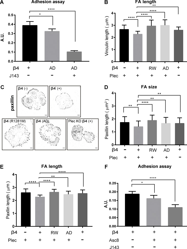
Figure S2.
Focal contact area varies depending on the expression and function of integrin β4. (A) Cells were treated with or without integrin α3–blocking mAb (J143; 20 µg/ml) in suspension before a short-term (45 min) adhesion assay was performed on a laminin-332–rich matrix substrate. Data are presented as the mean (± SD) from three independent experiments. *, P < 0.05; ****, P < 0.0001. (B) Quantification of FA length probed by vinculin with ImageJ. Data are presented as the mean (± SD) from three independent experiments, with ∼20 images per experiment. **, P < 0.01; ****, P < 0.0001. (C) Inverse black-and-white images of confocal micrographs of β4 (−), β4 (+), β4-R1281W, and β4-AD PA-JEB keratinocytes showing cell morphology and paxillin-stained FAs (black). Scale bars: 10 µm. (D and E) Quantification of FA size and length probed by paxillin with ImageJ. Data are presented as the mean (± SD) from two independent experiments, with ∼20 images per experiment. **, P < 0.01; ****, P < 0.0001. (F) Integrin β4 (+) PA-JEB keratinocytes were treated with integrin β4–blocking mAb (ASC-8; supernatant diluted 1:5) alone or together with integrin α3-blocking mAb (J143; 20 µg/ml). Data are presented as the mean (± SD) from three independent experiments. *, P < 0.05; ****, P < 0.0001.

