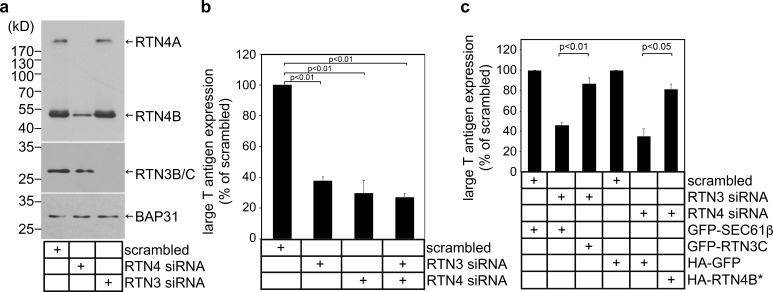
Figure 2.
RTN3 and RTN4 promote SV40 infection. (a) siRNA knockdown of RTN3 and RTN4. Cell extracts derived from CV-1 cells transfected with the indicated siRNA were subjected to SDS-PAGE and immunoblotting with the indicated antibodies. (b) CV-1 cells transfected with the indicated siRNAs were infected with SV40. 24 h after infection, cells were permeabilized, fixed, and stained for large T antigen. At least 1,000 cells were counted per condition over three biological replicates. (c) As in b, except cells were transfected with the indicated constructs 24 h before infection with SV40. Cells were then fixed, permeabilized, and stained for GFP/HA and large T antigen. Only cells expressing the GFP/HA construct were counted. At least 100 cells were counted per condition over three biological replicates. The graph represents the means ± SD. Student’s two-tailed t test was used to determine statistical significance.

