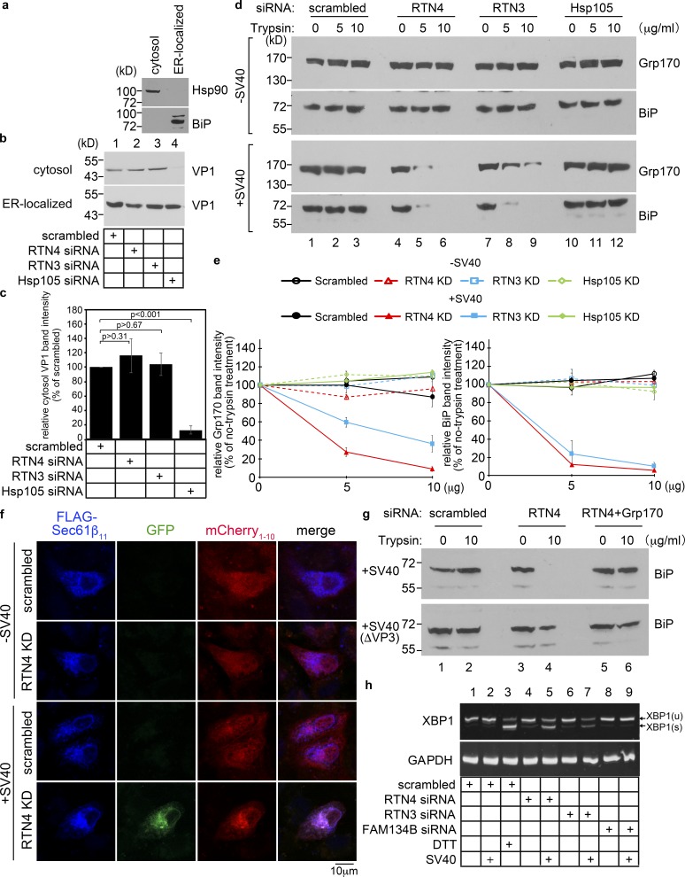
Figure 4.
RTN3 and RTN4 are essential to protect ER membrane integrity during SV40 infection. (a) CV-1 cells were semi-permeabilized with digitonin and centrifuged to generate the supernatant (cytosol) and pellet (membrane) fractions; material in the membrane fraction was further extracted by Triton X-100 to isolate ER-localized SV40. The cytosol and ER-localized fractions were analyzed for the presence of the cytosolic Hsp90 and the ER-resident BiP proteins. This procedure represents the ER-to-cytosol transport assay. (b) Cells transfected with the indicated siRNA were infected with SV40 for 16 h, and subjected to the ER-to-cytosol transport assay described in a. Both the cytosol and ER-localized fractions were analyzed by SDS-PAGE and immunoblotted with the indicated antibodies. (c) The intensity of the VP1 band in b was quantified by ImageJ using scans of films after ECL. The results represent the mean of three independent experiments. A two-tailed t test was used. Error bars, ± SD. (d) Protease protection assay to monitor the integrity of the ER membrane. CV-1 cells treated with the indicated siRNAs for 48 h were infected with SV40 for 16 h. Cells were subjected to semi-permeabilization as in panel a to generate the membrane fraction. This fraction (containing the ER membrane) was resolubilized in PBS with the indicated trypsin concentration. The sample was TCA-precipitated, and the precipitated material subjected to SDS-PAGE and immunoblotted with the ER luminal Grp170 and BiP antibodies. (e) Quantification of the percentage of Grp170 and BiP band intensity in d. Values represent means ± SDs from three independent experiments. (f) Split-GFP method to probe for the integrity of the ER membrane. COS-7 cells were treated with the indicated siRNAs for 24 h followed by cotransfection with FLAG-tagged Sec61β11 and cytosolic mCherry1-10. 24 h after transfection, cells mock-infected or infected with SV40 (MOI ∼100) for 16 h were fixed, stained with anti-FLAG antibody, and analyzed by a Zeiss LSM 800 confocal microscope. Scale bars, 10 µm. (g) CV-1 cells treated with the indicated siRNAs for 48 h were infected with 10 µg of WT SV40 or SV40(ΔVP3) for 16 h. Cells were subjected to the protease protection assay as in d. Cell extracts were collected, subjected to SDS-PAGE, and immunoblotted with an antibody against BiP. (h) CV-1 cells transfected with the indicated siRNAs for 2 d were mock infected or infected with SV40 (MOI ∼50) for 16 h. RNA was extracted and reverse-transcribed to cDNA, and the cDNA was amplified using PCR to identify splicing of the XBP1 mRNA. Cells treated with DTT served as a positive control.

