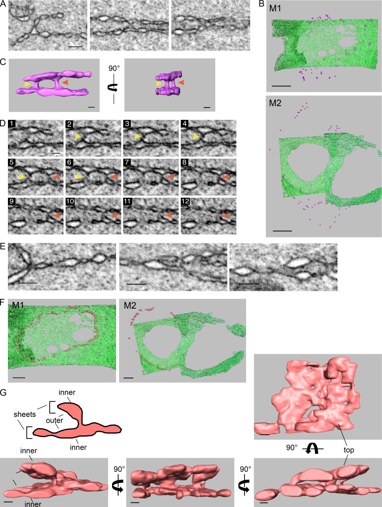
Figure 4.
At metaphase, the two pronuclei are connected by two types of junctions, outer–outer junctions and three-way sheet junctions, that surround the two-membrane interface. (A) 2D cross sections from FIB-SEM image volumes of outer–outer membrane junctions at the interface of metaphase pronuclei. Scale bar, 200 nm (applies to all panels). (B) Distribution of outer–outer junctions (purple spheres) relative the membrane interface (in green) of two metaphase one-cell embryos. Top panel, M1; bottom panel, M2. Scale bars, 1 µm. (C) Segmented volumes of two adjacent outer–outer junctions from the M1 metaphase embryo. The yellow and red arrowheads serve as markers for the two junctions shown in D. See also Fig. S2 A. Scale bars, 100 nm. (D) Consecutive FIB-SEM images of the outer–outer junctions shown in C. The distance between slices is 9 nm. See also Fig. S2 B. Scale bar, 100 nm (applies to all panels). (E) 2D cross sections from FIB-SEM image volumes of intersections between the four original pronuclear membranes and the two-membrane interface of the M1 embryo. Scale bars, 200 nm. (F) Distribution of the junctions (orange spheres) such as shown in E relative the membrane interface (in green) in two metaphase one-cell embryos. Left panel, M1; right panel, M2. Scale bar, 500 nm. (G) A segmented volume of three-way sheet junctions from embryo M2. Structures were rotated along the y axis as indicated, except for the right-most image, which is a top view of the segmented volume. See Fig. S2, C–E, for more examples. Scale bars, 100 nm.

