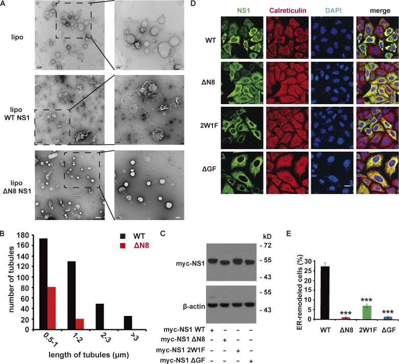
Figure 3.
ZIKV NS1 remodels membrane structure in vitro and in vivo. (A) EM imaging of liposomes (lipo) with or without NS1 proteins incubation. Liposomes (∼400 nm diameter) were incubated with NS1 proteins for 2 h at RT and then analyzed by EM after negative staining. Scale bar, left panel, 500 nm; right panel, 200 nm. (B) The length distribution of tubules induced by NS1 proteins. The length of tubules on liposomes was measured, and the number of tubules with different lengths was counted. (C) Expression of Myc-tagged WT or mutated ZIKV NS1 in HeLa cells. HeLa cells were transfected with WT or mutated ZIKV NS1. At 24 h after transfection, NS1 was detected by Western blotting with an anti-Myc antibody. (D) HeLa cells expressing Myc-tagged WT or mutated NS1 were stained with antibodies against Myc-tag (green) or calreticulin (red). Scale bar, 20 µm. (E) Percentage of cells with ER remodeling mediated by WT or mutated NS1. The percentage is defined as the ratio of cells with ER remodeling to NS1-expressing cells. The data are the mean ± SEM. The P values are obtained from a two-tailed t test. ***, P < 0.001.

