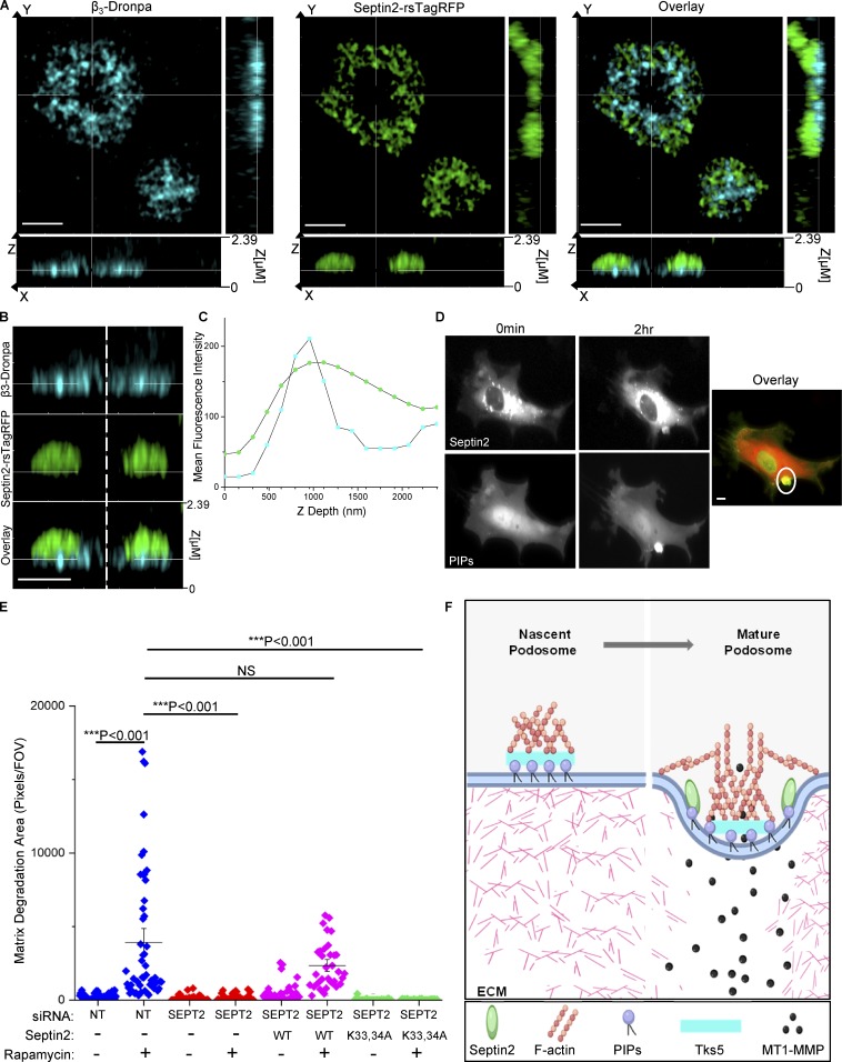
Figure 5.
Septin2 binding to PIPs mediates podosome function. (A–C) Substrate-proximal localization of Septin2 in podosomes depicted by Airyscan confocal imaging. HPAECs expressing Septin2-rsTagRFP and β3-Dronpa, transduced with adenoviral constructs for expression of RapR-Src and FRB and fixed following 0.5 h of Src activation. (A) Orthogonal view of Src-induced rosette (scale bar = 3 µm). (B) Zoomed-in view of orthogonal slice as in A (scale bar = 2 µm). (C) Depth profile of β3 integrin (blue) and Septin2 (green) localization in the Z axial direction from the rosette depicted in A. (D) HPAECs expressing Septin2-mCherry (top) and a Venus-fused PIP binding probe (bottom) were imaged live using widefield illumination before and after Src activation (scale bar = 5 µm). Colocalization of Septin2 (red) and PIP binding probe (green) is indicated by the white circle in the overlay panel. (E) Rescue of matrix degradation by exogenous expression of Septin2. HPAECs were treated with siRNA for Septin2 (SEPT2) or control siRNA (NT) for 72 h. Cells were transduced with adenoviral constructs for expression of RapR-Src and FRB and with lentiviral constructs for expression of either WT mouse Sept2-mCherry or its mutant (Lys33Ala/Lys34Ala, K33,34A). Total matrix degradation was measured from at least 10 fields of view per condition from three to five independent experiments. Individual data points are shown around mean value ± SE (whiskers). Significance was determined using a two-sample t test (comparisons indicated by lines; FOV, field of view). (F) Model depicting the role of Septin2 in podosome maturation. Podosome initiation (left panel): Tyrosine kinase Src is activated downstream of growth factor signaling. Src signals through multiple intermediates including PI3K, which generates PIPs. These PIPs localize to sites of podosome adhesion and bind scaffolding protein Tks5. Podosome maturation (right panel): Septin2 localizes to podosomes downstream of Src activation and binds PIPs via its polybasic domain. The Septin cytoskeleton creates a confinement border around the adhesion ring, allowing for maturation and localized matrix degradation (image created with BioRender).

