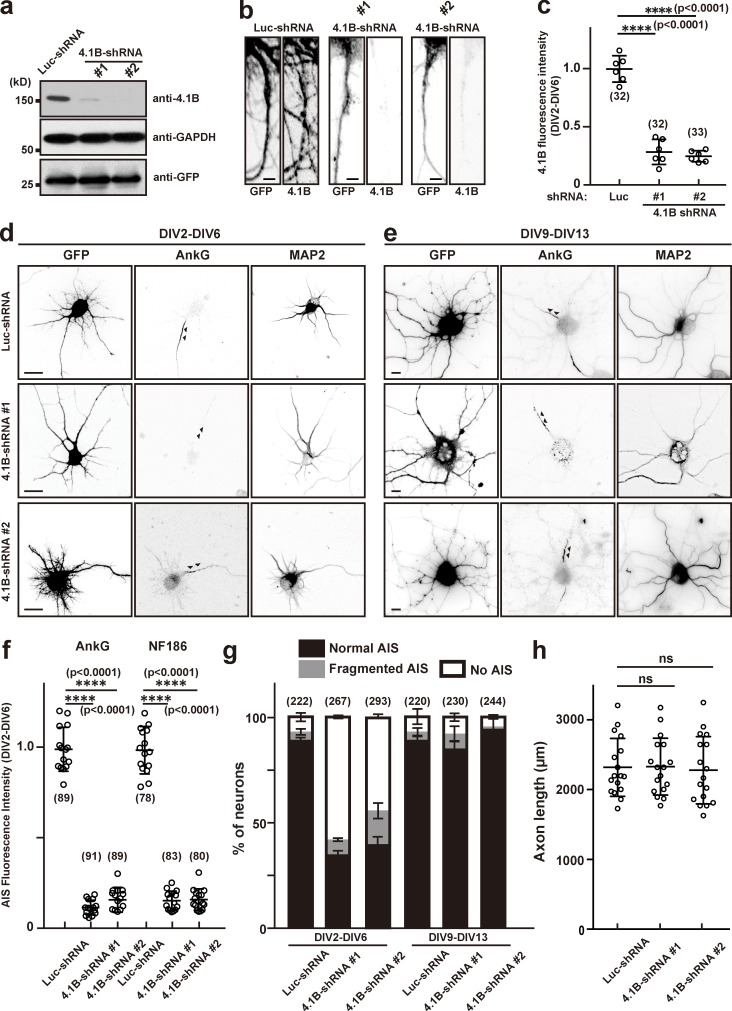
Figure 6.
Silencing expression of protein 4.1B disrupts AIS assembly but not maintenance. (a) Mouse neuroblastoma Neuro-2a cells were transfected with an shRNA construct for control, (Luc-shRNA), 4.1B-shRNA #1, or 4.1B-shRNA #2 to validate shRNA efficiency. The expression levels of 4.1B, GAPDH, and GFP were determined by immunoblotting. (b) Immunostaining of hippocampal neurons transfected with Luc-shRNA, 4.1B-shRNA #1, or 4.1B-shRNA #2 at DIV2 and stained at DIV6. Scale bars = 3 µm. (c) Quantification of 4.1B immunofluorescence in hippocampal neurons after knockdown by shRNA. Mean ± SEM is shown (****, P < 0.0001; n = 6 independent experiments, repeated measures one-way ANOVA; the number of neurons analyzed for each condition is also shown). (d and e) Hippocampal neurons were transfected with Luc-shRNA, 4.1B-shRNA #1, or 4.1B-shRNA #2 at DIV2 and stained at DIV6 (d), or transfected at DIV9 and stained at DIV13 (e) using antibodies against GFP, AnkG, and MAP2. Arrowheads indicate AIS in hippocampal neurons. Scale bars = 5 µm. (f) Fluorescence intensity of AnkG and NF186 at the AIS in neurons transfected using Luc- or 4.1B-shRNAs; neurons were transfected at DIV2 and analyzed at DIV6. Mean ± SEM is shown (****, P < 0.0001; n = 15 independent experiments, repeated measures one-way ANOVA; the number of neurons analyzed for each condition is also shown). (g) Quantification of the staining pattern for AnkG in shRNA-transfected neurons. The number of neurons counted is shown. Mean ± SEM is shown. No statistical comparison was performed. (h) Axon length in neurons transfected with control or 4.1B-shRNAs. Mean ± SEM is shown (N = 3 independent experiments, n = 17 total cells measured; one neuron was measured per coverslip; repeated measures one-way ANOVA).

