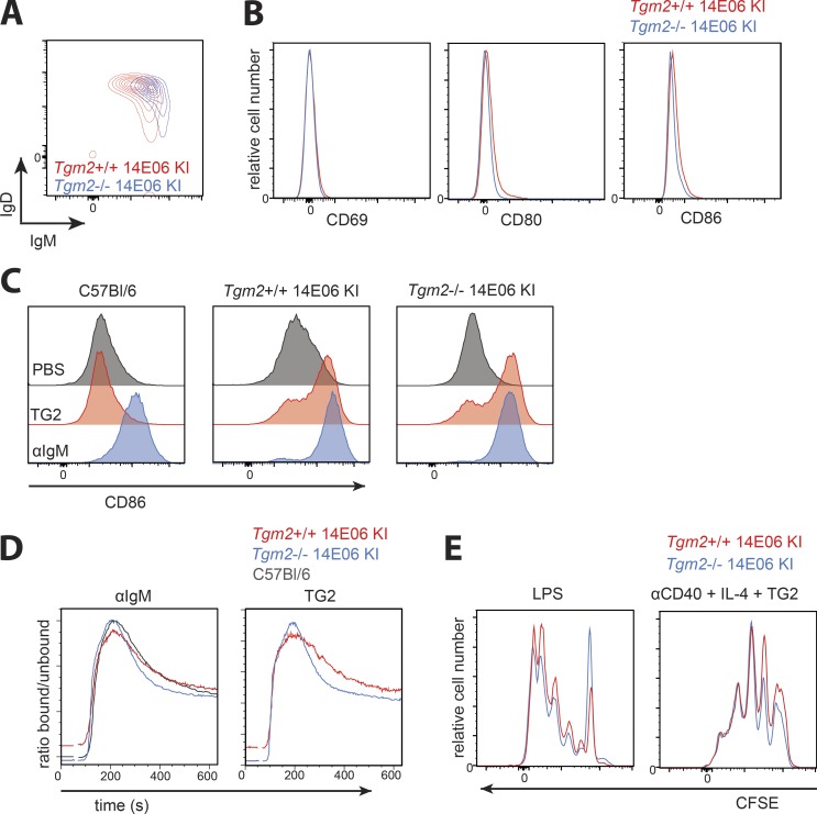
Figure 3.
14E06 Ig KI B cells are not anergic. (A) Representative plot of spleen B220+ B cells of Tgm2+/+ (red) and Tgm2−/− 14E06 KI mice (blue) stained for anti-IgM and anti-IgD. (B) Histogram plots gated on B220+ TG2-tetramer+ B cells isolated from spleens of Tgm2+/+ (red) and Tgm2−/− (blue) 14E06 KI mice that were stained for CD69, CD80, and CD86. (C) Induction of CD86 after 18 h culture of isolated B cells from spleens of C57Bl/6, Tgm2+/+ 14E06 KI, and Tgm2−/− 14E06 KI mice in the presence of 2 µg/ml TG2 or 10 µg/ml anti-IgM. (D) Ca2+ flux of C57Bl/6, Tgm2+/+ 14E06 KI, and Tgm2−/− 14E06 KI B cells. Splenic B cells were loaded with the intracellular calcium indicator Indo-1. Baseline Ca2+ levels were acquired for 30 s before the cells were stimulated with 10 µg/ml anti-IgM or 2 µg/ml TG2. (E) CFSE dilution after 72 h of culture of isolated B cells of Tgm2+/+ (red) and Tgm2−/− (blue) 14E06 KI mice stimulated with 2 µg TG2 in the presence of 5 µg/ml anti-CD40 and 50 ng/ml IL-4 or 5 µg/ml LPS. All data are representative of at least three independent experiments (A–E). For C57Bl/6 control mice, we used either commercially obtained C57Bl/6 from Janvier Laboratoires or nontransgenic littermates from in-house breedings.

