Figure 6.
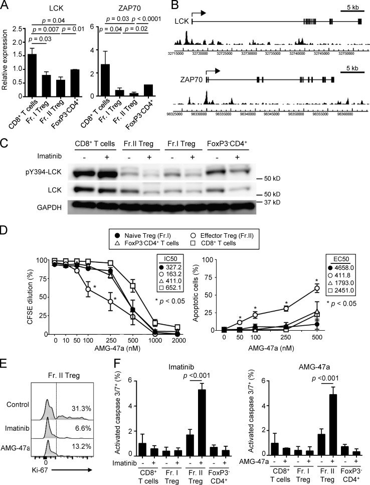
Imatinib inhibits LCK and selectively causes apoptosis in T reg cells. (A) LCK and ZAP-70 mRNA expressions in naive CD8+ T cells, Fr. I naive T reg cells, Fr. II eT reg cells, and naive CD4+ T cells prepared from PBMCs of healthy donors. LCK (n = 6) and ZAP-70 (n = 5) mRNA levels relative to GAPDH were measured by quantitative real-time PCR. Data are pooled from more than two independent experiments. (B) FoxP3 binding near the promoter regions of human LCK and ZAP-70 genes. The arrows indicate transcription start sites, boxes indicate exons, and peaks indicate FoxP3-binding sites from SRA data analysis (SRX060160; Birzele et al., 2011) by Integrative Genomics Viewer software. (C) Total and phosphorylated LCK (pY394-LCK) protein expression measured by Western blotting. Each cell population sorted from PBMCs of healthy donors was incubated with or without 10 µM imatinib for 1 h (n = 5). Data are representative of five independent experiments. (D) Proliferation inhibition and apoptosis induction by the LCK inhibitor AMG-47a. Subsets of CD4+ T cells (Fr. I and II and FoxP3−CD4+ T cells) and CD8+ T cells prepared from PBMCs of healthy donors (n = 3) were stimulated with anti-CD3 and anti-CD28 mAbs with or without graded doses of AMG-47a for 5 d. Proliferation was assessed by CFSE dilution, and apoptosis was assessed by Annexin V and 7-AAD staining. IC50 for proliferation and EC50 for apoptosis are shown in the boxes. Data are pooled from more than two independent experiments. (E) Representative staining (n = 3 each) of Ki-67 in Fr. II eT reg cells from PBMCs of healthy donors treated with 10 µM imatinib or 250 nM AMG-47a for 4 d with anti-CD3 and anti-CD28 mAbs. Percentages of Ki-67+ cells were indicated. Data are pooled from more than two independent experiments. (F) The frequencies of cells with activated caspase 3 and 7, prepared from PBMCs of healthy donors, after treatment with or without 10 µM imatinib or 250 nM AMG-47a for 4 d with anti-CD3 and anti-CD28 mAbs. Caspase 3/7+ cells negative for SYTOX AADvanced cell impermeant nucleic acid stain were measured (n = 3 each). Data are pooled from more than two independent experiments. Error bars indicate means ± SEM in A, D, and F. Statistical significance was assessed by Student’s two-tailed paired t test in A and two-way ANOVA with Holm-Sidak multiple comparisons to CD8+ T cells in D, or to imatinib or AMG-47a–treated Fr. II T reg cells in F.

