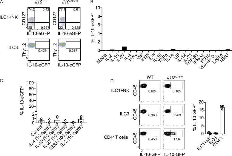
Figure S1.
IL-10 production in NK cells, ILC1s, and ILC3s. (A) Expression of IL-10–eGFP in NK1.1+ cells (pooled ILC1s and NK cells) and ILC3s isolated from naive small intestine lamina propria. (B) Screen for IL-10 elicitors using sorted small intestine ILC3s cultured for 2 d with IL-23, IL-1β, IL-7, and a candidate mediator. (C) Expression of IL-10–eGFP in small intestine lamina propria ILC3s sorted from biological replicates and cultured for 2 d with IL-23, IL-1β, IL-7, and molecules that were found to induce IL-10 in ILC2s (n = 4). (D) Expression of IL-10–eGFP in NK1.1+ cells (pooled ILC1s and NK cells), ILC3s, and CD4+ T cells isolated from small intestine lamina propria of mice 4 d after inoculation with 108 CFU of C. difficile VPI 10463 (n = 3). Bars indicate means ± SD. Data from A, C, and D are representative of two independent experiments.

