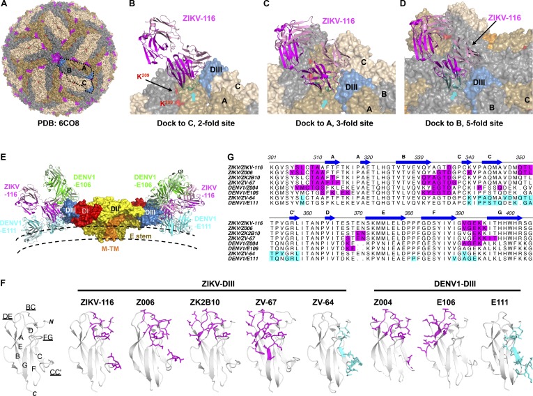
Figure 7.
Epitope comparison of neutralizing DIII-specific mAbs against ZIKV and DENV1. (A) Mapping of the ZIKV-116 epitope onto the mature ZIKV virion (PDB 6CO8). E proteins in three symmetries are colored in wheat (twofold; C), olive (threefold; A), and gray (fivefold; B). The ZIKV-116 epitope is colored in magenta, and K209(ZIKV) correlating to R/K204(DENV1) is colored in red. (B–D) Docking of the ZIKV-116 Fab onto representative twofold (B), threefold (C), and fivefold (D) sites. Clashes with adjacent E proteins were highlighted in cyan and indicated with arrows. (E) Docking of the ZIKV-116, DENV1-E106 (exposed A-strand epitope), and DENV1-E111 (cryptic CC′-loop epitope) Fabs onto the M-E dimer of the mature virion. ZIKV-116 binding to the LR of DIII is located between the positions of DENV1-E106 and DENV1-E111. (F) Ribbon diagrams of DIII, and epitopes recognized by mAbs are rendered as sticks. LR/A-strand epitopes are colored in magenta, and CC′-loop epitope is colored in cyan. (G) Sequence alignment of ZIKV and DENV1 with highlighted antibody epitopes (same coloring as in F). ZIKV-Z006 (PDB 5VIG), ZIKV-ZK2B10 (PDB 6JEP), ZIKV-ZV-67 (PDB 5KVG), DENV1-Z004 (PDB 5VIC), DENV1-E106 (PDB 4L5F), ZIKV-ZV-64 (PDB 5KVF), and DENV1-E111 (PDB 4FFY) were used for the analysis.

