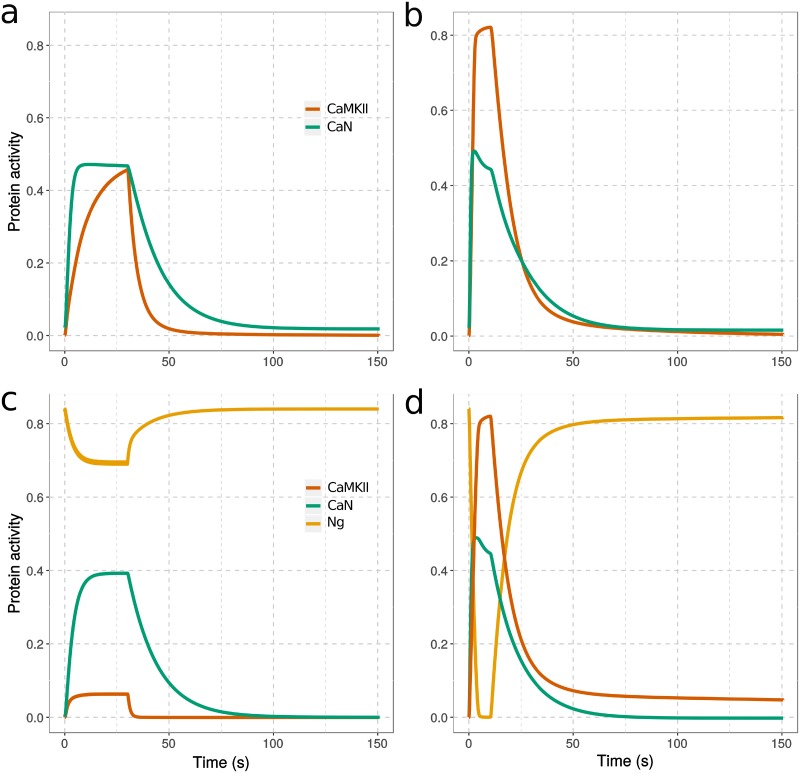
Fig 7. Change of protein activity in response to calcium spikes.
Simulations in absence (a,b) or presence (c,d) of Ng, from equilibrium and during stimulation by 300 calcium spikes at 10 Hz (a,c) and 30 Hz (b,d). The protein activities were defined as follow: fraction of CaMKII monomers bound to CaM and/or phosphorylated multiplied by CaMKII kcat for GluR1, fraction of CaN bound to CaM multiplied by CaNA kcat for GluR1, fraction of Ng bound to CaM. [CaM]tot = 40 μM, [CaMKII]tot = 80 μM, [CaN]tot = 8 μM, and [Ng]tot = 40 μM; kcatCaMKII = 2 s-1, kcatCaN = 0.5 s-1. All the concentrations of perspective CaM binding proteins are assigned according to the proteomic study in the hippocampus CA1 region [64].

