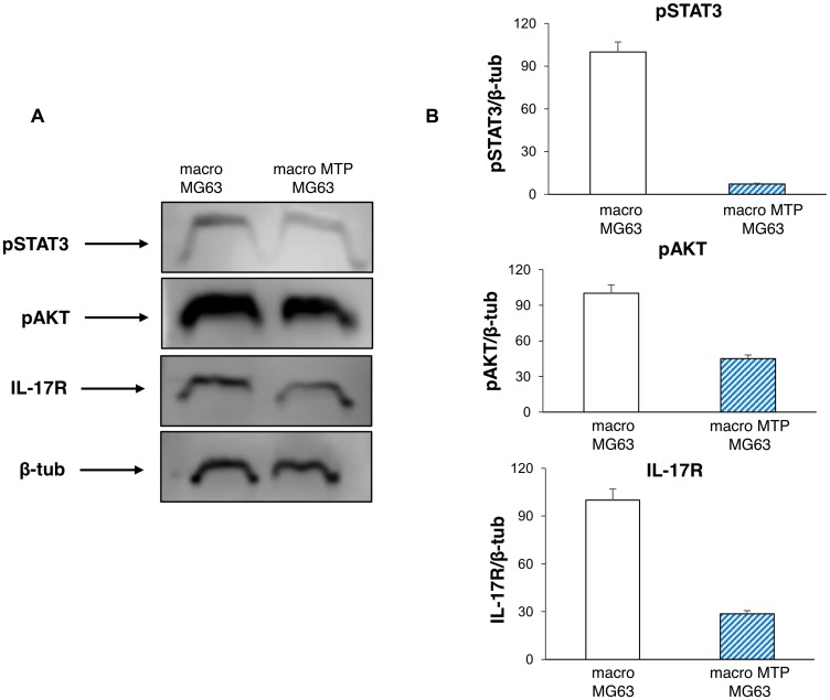
Figure 6. Effects of Mifamurtide (MTP) on proliferation and invasive properties of MG63 co-cultured with macrophages.
(A) pSTAT3, pAKT, IL-17R protein expression levels in MG63 co-cultured with macrophages activated or not with MTP [100 µM] determined by Western Blot, starting from 15 µg of total lysates. The most representative images are displayed. The proteins were detected using Image Studio Digits software and the intensity ratios of immunoblots compared to macrophages-MG63 co-culture taken as 100 (arbitrary unit), were quantified after normalizing with respective loading controls for the housekeeping protein β-Tubulin. (B) The graphs represent the relative quantification for pSTAT3, pAKT, IL-17R expression as mean ± SD of three independent experiments. A t-test has been used to evaluate the statistical differences in protein expression levels. *indicates p ≤ 0.05 compared to macrophages-MG63 co-culture.

