Figure 5.

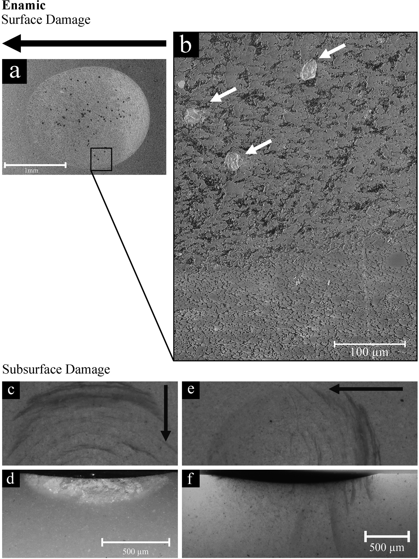
Surface and subsurface damage in Enamic after 104 cycles. Black arrows indicate the direction of sliding. An important surface degradation is observed inside the wear scar as observed in the SEM image a and its inset b. Partially removed feldspathic particles can be observed inside the crater (white arrows) still embedded in the polymer network. The fracture and removal of the glassy matrix leaves a porous and rough surface, which contrasts strongly with the compact and polished appearance of the material surrounding the crater. The same surface features are observed throughout the crater. Partial cone (herringbone) cracks were visible in the direction of sliding under transilluminated light in the stereomicroscope, as depicted in c and e. A higher density of cracks is observed at the tail of the indenter path. In d, a transversal section of one of this partial cone cracks is presented. The sagittally-sectioned sample depicted in f shows the series of partial cone cracks penetrating deep into the material. Note the steep penetration angles of the cracks, ranging between 75° and 85°. A higher magnification was used for images c and d, than for e and f.
