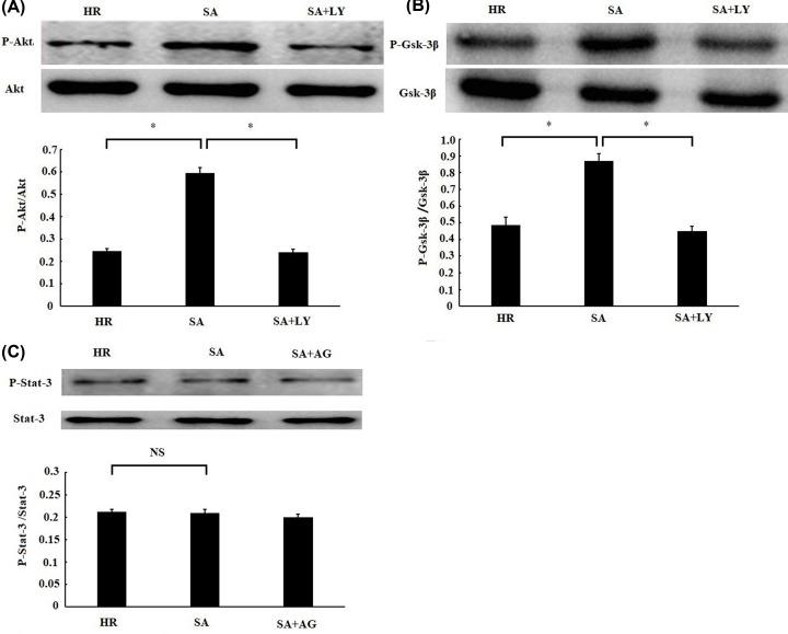
Figure 5. Sappanone A (SA) pretreatment activated PI3K–Akt–Gsk-3β signal pathway.
H9c2 cardiomyocytes were treated by 25 µM SA for 1 h, followed by 6 h of hypoxia/3 h of reoxygenation. About 10 μM LY294002 (LY), a PI3K inhibitor, was given 1 h prior to SA treatment. About 10 μM AG490 (AG), a STAT3 inhibitor, was given 1 h before SA treatment. The phosphorylation levels of Akt (A), Gsk-3β (B), and Stat-3 (C) were detected by Western blotting. Data are presented as the mean ± standard deviation from three independent experiments. * P < 0.05, NS, no significance.

