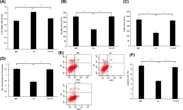
Figure 6. The cardioprotection of Sappanone A (SA) was abrogated by inhibition of PI3K/Akt.
H9c2 cardiomyocytes were treated by 25 µM SA for 1 h, followed by 6 h of hypoxia/3 h of reoxygenation. About 10 μM LY294002 (LY), a PI3K inhibitor, was given 1 h prior to SA treatment. (A) Cell viability was detected by CCK-8 assay. (B) The creatine kinase-MB (CK-MB) and (C) lactate dehydrogenase (LDH) activity in culture medium were measured by spectrophotometry. (D) The concentration of Troponin (cTnI) in culture medium was measured by spectrophotometry. (E) Cell apoptosis was detected by flow cytometry. Cells in the lower right quadrant represent apoptosis cells. (F) Statistical analysis of apootosis rate. Data are presented as the mean ± standard deviation from three independent experiments; * P < 0.05.

