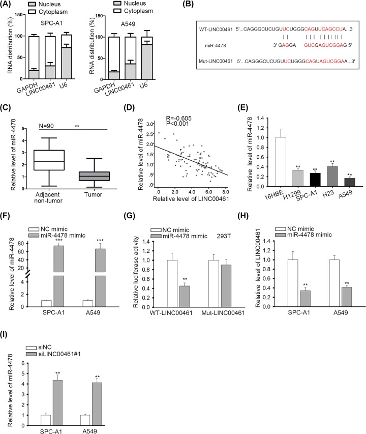
Figure 2. LINC00461 sponged miR-4478 in NSCLC cells.
(A) Cellular location of LINC00461 in SPC-A1 and A549 cells was analyzed by subcellular fractionation. (B) The binding site between LINC00461 and miR-4478 was predicted and the mutant site was constructed. (C) RT-qPCR was applied for detecting miR-4478 expression in NSCLC tissues and matched para-cancerous tissues. (D) Spearman’s correlation curve showed the negative relation between miR-4478 and LINC00461. (E) RT-qPCR exhibited miR-4478 expression in NSCLC cells. (F) Results of RT-qPCR confirmed the overexpression efficiency of miR-4478 mimic. (G) The luciferase reporter assay validated the interaction between LINC00461 and miR-4478. (H) Effect of miR-4478 mimic on LINC00461 expression was confirmed. (I) MiR-4478 expression in siLINC00461 transfected cells was detected by RT-qPCR. **P<0.01, ***P<0.001.

